 (515) 280-3100
Critical case of Respiratory Distress
Written by: Becky L. • 2016 Scholar
 Tigger Goddard, a four year old, neutered male, domestic short hair presented to Iowa Veterinary Specialty through the emergency department for respiratory distress. Previously Tigger had presented at his primary care veterinarian where chest x-rays where taken and a dose of Lasix, a strong diuretic agent, was given. Upon presentation at IVS, Tigger had a low body temperature (98.6 F), was tachycardic (a high heart rate of 200bpm) and had an increased respiratory rate with increased abdominal effort.
His pulse oximetry was at 98% with room oxygen levels, meaning he was still able to oxygenate well but with increased effort. Initial blood work showed a leukocytosis (high white blood cell count) at 22,000 and mature neutrophilia ( a certain type of white blood cell) at 19,000, hyponatremia (low sodium), and was FeLV and FIV negative. Thoracic radiographs showed consolidation of the right and left cranial lung lobes and the cranial and dorsal margins could not be visualized. A small amount of pleural effusion (fluid in the chest between the chest wall and the lungs) was noted on the right side with pleural fissure lines noted on the left. With the results of his blood work and his presenting clinical signs, hospitalization with IV fluids and antibiotics were recommended as well as a thorax ultrasound for further diagnostics. While there are many causes for respiratory distress in cats, Tigger’s diagnosis included suspect infectious pneumonia with possible pyothorax as well as pulmonary neoplasia or cardiogenic pulmonary edema. A more accurate diagnosis would require a thoracic ultrasound and a sample of the fluid in his chest in order to determine if the fluid was due to an infection, to heart disease or cancer.
Tigger was hospitalized and placed in an oxygen cage on fluids with a broad spectrum antibiotics, Unasyn and Baytril, as well as nebulization and coupage to help him breath better.
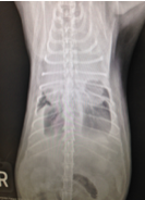 While still on treatments for his increased respiratory effort, Tigger continued to have trouble breathing and had decreased lung sounds in all fields. While re-check radiographs did show an improvement in his lung filling capacity, there was still fluid in his chest.
There are different types of fluid that can be found in the chest such as blood, pus or lymph fluid. These fluids are characterized as being an exudate, modified transudate or pure transudate based on their cellular and protein composition. A thoracentesis is an emergency procedure that can be diagnostic and therapeutic. It allows the clinician to drain the fluid from the chest in order to perform more diagnostic tests on the fluid and to determine the source of the fluid. Actively removing the fluid from the chest also allows the lungs to more fully and easily expand and allows immediate relief for the patient. Unfortunately it is not a curative procedure since the fluid can build back up again if the underlying medical condition is not found and treated.
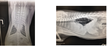
In order to help Tigger breath better, he underwent a thoracocentesis (removing some of the fluid with a needle and syringe). He was maintained on flow by oxygen during the procedure while a 20G needle was placed in his thorax and 108 ml of slightly cloudy yellow to clear fluid was removed from his right side and 89 ml of clear to pink fluid was removed from his left. After the procedure, Tigger’s respiratory rate did not decrease but he no longer had increased abdominal breathing effort. Post procedural radiographs showed improvement in his lung capacity but unfortunately there was still a soft tissue opacity in the anterior lung fields suggestive of a mediastinal mass (a tumor in the space in the center of the chest). Treatment options for the new finding would include surgery with possible chemotherapy.
The most common causes of anterior mediastinal masses in small animals are lymphomas and thymomas. In cats 1-3 years old, lymphoma tends to be the most common finding while thymomas are more common in cats older than 8 years. Other non-neoplastic (noncancerous) causes of anterior mediastinal masses are thymic or mediastinal hematomas and ultimobranchial cysts.
Despite the thoracocentesis, Tigger’s respiratory efforts continued to be increased and with a guarded prognosis, the owner’s consented to euthanasia. Both IVS clinicians and Tigger’s owners were able to provide the best supportive and diagnostic care in order to find Tigger’s underlying medical condition. Considering his guarded prognosis and declining clinical signs, euthanasia was the human method to end his suffering.
References:
- Couto G. G, Nelson E. R., Small Animal Internal Medicine fifth Ed.,Elsevier Mosby.2014. 1154-1157
- Silverston C. D, Hopper K., Small Animal Critical Care Medicine. Saunders Elsevier. 2009., 131-133.