
Feline Femoral Head Fracture
Written by Molly Hoffenberg • 2025 Scholar
Signalment & History
Dorito is a 1-year-old neutered male Domestic Shorthair who presented to IVS unable to use his back legs. The owner reported hearing the patient vocalizing loudly in another room, after which Dorito was found non-weight-bearing on his hind limbs.
Physical Exam Findings & Diagnostics
On presentation, Dorito was weight-bearing on three limbs and toe-touching on the left pelvic limb. He exhibited discomfort upon manipulation of the left femur, and moderate crepitus was palpated at the level of the left coxofemoral joint under sedation. All vital parameters were within normal limits. Neurologic examination was appropriate, and body condition score (BCS) was 6/9. Dorito was deemed clinically stable but painful, with a pain score of 3/4.
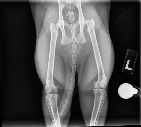
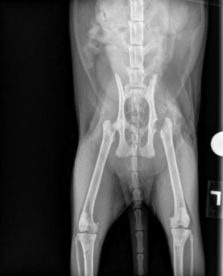
Radiographs of the pelvis and left femur were performed and reviewed by the IVS on staff veterinarian (Dr. Blong) and an IDEXX board-certified radiologist. Imaging revealed a complete, transverse Salter-Harris type I fracture of the left femoral head physis along with delayed physeal closure. This indicated that the fracture occurred at the growth plate and caused a slipped epiphysis.
Diagnosis
Based on clinical findings and radiographic interpretation, Dorito was diagnosed with a left femoral head fracture.
Treatment & Outcome
Surgical intervention was recommended. Dorito was managed overnight on medical therapy, including a single dose of transdermal buprenorphine (Zorbium, 20 mg/mL, 1 mL), oral gabapentin (100 mg PO q8h), and strict crate rest pending surgical consultation.
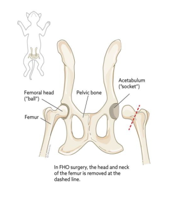
The following day, Dorito underwent a femoral head ostectomy (FHO) under the care of Dr. Law at IVS. A craniolateral approach to the left coxofemoral joint was performed. The incision was centered over the greater trochanter, with retraction of the middle and deep gluteal muscles caudally to expose the femoral head and neck. The joint capsule was incised to facilitate luxation and external rotation of the femoral neck, enabling visualization of the fractured epiphysis. The femoral neck was osteotomized from the greater to the lesser trochanter. The femoral head was removed using towel clamps. The ostectomy site was smoothed, and the limb was manipulated through a range of motion to confirm no residual rough bony edges. Additional ostectomy of the lesser trochanter was performed. The gluteal muscle, quadriceps, and tensor fasciae latae were re-apposed, and the surgical site was closed in subcutaneous and cutaneous layers.
Dorito tolerated anesthesia and surgery well, and his pain was well-controlled postoperatively. He was managed in-hospital with IV cefazolin, Zorbium, gabapentin, and robenacoxib (Onsior). Dorito recovered uneventfully and was discharged with oral gabapentin and Onsior, along with instructions for strict confinement and activity restriction for three weeks.
Femoral Head Fracture in Young Male Neutered Cats
Spontaneous capital physeal fractures are commonly seen in young (typically under two years of age), overweight, neutered male cats with no history of trauma. It is well proven that neutering delays physeal closure in male cats, as the sex hormones, particularly testosterone and estrogen, play a critical role in cartilage maturation and growth plate fusion. Following neutering, the reduction in circulating sex hormone levels results in delayed physeal closure, predisposing the immature cartilage to fracture, even in the absence of external trauma.
A similar condition, metaphyseal osteopathy, also tends to affect young, overweight, neutered male cats and can present with clinical signs that closely resemble those of spontaneous capital physeal fractures. Radiographically, this condition is characterized by severe bone lysis and remodeling at the femoral neck, often accompanied by fractures through the capital physis. Histopathologic findings may include focal irregularities, thickening, and fragmentation of the articular cartilage. While the exact pathogenesis remains unclear, it is hypothesized that altered vascular supply to the metaphysis—whether increased or decreased—may lead to ischemia, necrosis, structural collapse, and subsequent fracture of the femoral neck.
Treatment options for femoral head fractures, whether spontaneous or secondary to suspected metaphyseal osteopathy, vary depending on the severity and chronicity of the lesion. One surgical approach is open reduction and internal fixation using Kirschner wires (K-wires), which aims to preserve the native anatomy and function of the coxofemoral joint. Outcomes for this technique are generally favorable, with most cats achieving good to excellent functional recovery. Alternatively, a femoral head ostectomy (FHO procedure) may be performed. This procedure involves removing the head of the femur, allowing the surrounding muscles to hold the femur in place. This creates a false joint but allows free mobility of the coxofemoral joint without the typical ball-and-socket joint. While this procedure is also associated with good to excellent outcomes, in many cases potential complications include persistent lameness, limb shortening, reduced range of motion, or discomfort upon manipulation of the joint.
Although surgical intervention is typically recommended, conservative management may be considered in select cases—particularly in young cats presenting with acute, non-displaced fractures and no radiographic evidence of bone lysis or remodeling. Conservative therapy consists of strict exercise restriction combined with an analgesia in an effort to promote natural healing. However, if metaphyseal osteopathy is suspected, conservative treatment alone is unlikely to resolve clinical signs due to the underlying pathology.
Sources
- Lafuente P. (2011). Young, male neutered, obese, lame? Non-traumatic fractures of the femoral head and neck. Journal of feline medicine and surgery, 13(7), 498–507. https://doi.org/10.1016/j.jfms.2011.05.007
- Schwartz G. (2013). Spontaneous capital femoral physeal fracture in a cat. The Canadian veterinary journal = La revue veterinaire canadienne, 54(7), 698–700.
- Weir, M., & Barnette, C. (2022). Femoral head ostectomy (FHO) in cats: VCA Animal Hospitals. Vca. https://vcahospitals.com/know-your-pet/femoral-head-ostectomy-fho-in-cats