
Foreign Body – Rocky Ingestion
Written by Abby Stewart • 2024 Scholar
Presentation
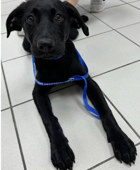
Casey is a 5 month old, intact, female black labrador puppy. She presented to Iowa Veterinary Specialties this evening for being restless, pacing and refusing to lay down. She has been eating, drinking, urinating, and defecating normally. Patient did receive vaccines about two days prior to presentation. Upon presentation she was full of energy and very curious about everything in
the clinic.
Physical Exam
- Weight: 12.1 kg
- Heart Rate: 180 bpm (Normal: 60-160)
- Respiratory Rate: 60 bpm (Normal: 10-40)
- MM/CRT: pink, <2 seconds (Normal: Pink, <2 seconds)
- Temperature: 102.8 degrees Fahrenheit (Normal: 100-102.5)
- BCS: 5/9
- Physical Exam: Fairly unremarkable. Abdomen was soft and non-painful with no appreciable masses. Neurologically/Mentally appropriate. No heart arrhythmias auscultated, and normal bronchovesicular sounds. Casey’s heart rate, respiratory rate, and temperature were slightly elevated, but this was likely due to her excitement from being at the clinic, and thus not extremely concerning at this time.
Differentials
- Urinary tract infection (UTI)
- Systemic illness/infection
- Abdominal pain (GI issues) – constipation, food bloat, foreign body, etc…
- Cytokine reaction from vaccines
Diagnostic Tests
To attain more information, abdominal radiographs (X-rays), blood work including a CBC and a chemistry panel, and a urinalysis were recommended. The owner approved all diagnostics. Due to the findings obtained on radiographs, the urinalysis was not performed.
Blood work findings:
Casey’s blood work returned a mild microcytic, normochromic non-regenerative anemia. Evidenced by a slightly low hematocrit (HCT), hemoglobin (HGB), and mean corpuscular volume (MCV). There was no evidence of reticulocytosis, indicating that the anemia was non-regenerative. This is not an uncommon finding in young patients, due to their limited iron stores.
She also had a lymphocytosis, most likely due to her young age and excitement resulting in the epinephrine response. When epinephrine is released, it essentially causes a redistribution of lymphocytes from the marginated lymphocyte pool to the circulating lymphocyte pool, resulting in an increased lymphocyte count on blood work.
All other values were within normal limits. Anemia is defined as a decrease in HCT, HGB, and/or red blood cells (RBC). A lymphocytosis is an increase in the number of lymphocytes.
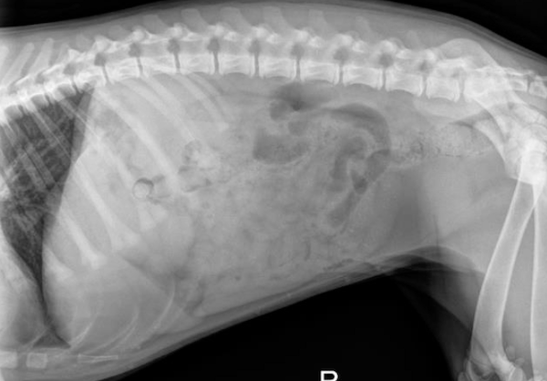
Radiograph findings:
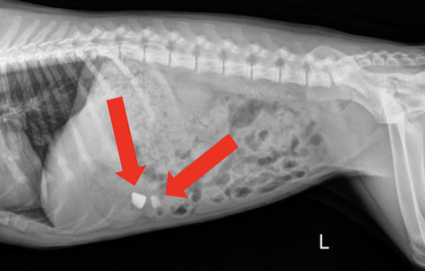
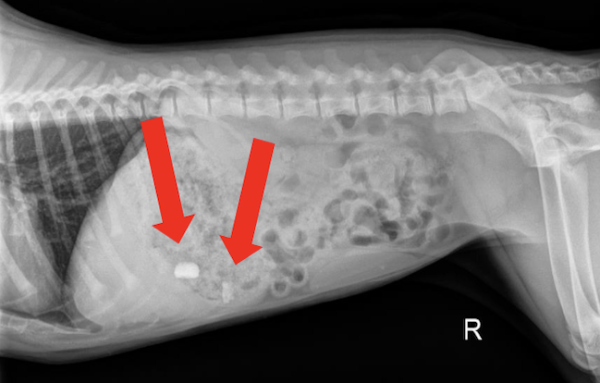
The radiographs returned two radiopaque foreign materials in the stomach (shown above by the red arrows). These objects were suspected to be rocks. In order to confirm the location of these objects two views were taken including the lateral views above as well as a ventrodorsal (VD) view (not pictured).
Diagnosis
Foreign bodies in the stomach. One large rock, and one smaller rock near the pylorus. The smaller rock was more concerning due to its location at the pylorus and the potential for it to become lodged at this location or pass into the duodenum.
Treatment
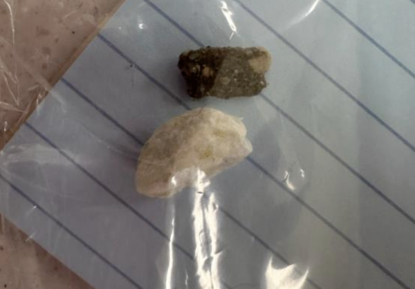
In order to remove the two rocks in the stomach, emesis was induced using 0.04 mg/kg apomorphine intravenously. Apomorphine is a synthetic opiate that works as a dopamine-2 receptor agonist and induces emesis in canines. Casey did produce vomitus that contained both rocks. Upon successful removal of the rocks from her stomach, Casey was given 1 mg/kg Cerenia (Maropitant citrate) via subcutaneous injection. Cerenia is a neurokinin-1 receptor antagonist that blocks substance P leading to antiemesis effects. Repeat abdominal radiographs were taken to confirm all rocks had been removed from her stomach. She was then discharged to return home.
Other Potential treatments
Had Casey failed to produce the two rocks, endoscopy or an abdominal exploratory with a gastrostomy would have been recommended to remove the foreign bodies in the stomach. A brief description of each procedure is described below.
- Endoscopy: An endoscopy is a minimally invasive, non-surgical procedure performed using an endoscope instrument. The endoscope instrument has a viewing port and/or video camera attached to it. In this case a flexible endoscope would have been inserted through the mouth, the esophagus, stomach and possibly into the duodenum. As the instrument was inserted through these organs, each organ would have been examined for irritation, inflammation and other abnormalities. In addition to examining these organs, the rocks could have been retrieved from the stomach using the endoscope instrument and removed without any surgical incisions. Anesthesia is required for this procedure. If endoscopy was unsuccessful in removing the rocks, then surgery would be warranted to remove them.
- Abdominal Exploratory + Gastrotomy: In this surgery the abdomen is incised and all structures of the abdominal cavity are examined/palpated to look for lesions and abnormalities. Then, in this case the stomach would have been palpated for the rocks. The stomach would have been isolated, packed off with laparotomy sponges, and incised. The foreign material would be retrieved and the stomach would be closed. The abdominal cavity would then be closed and the surgery concluded. The animal would have been sedated for this procedure.
References & Citations
- Atilla, A. (2017). Gastrointestinal surgery – some tips and tricks. Companion animal, 22(6). https://doi.org/10.12968/coan.2017.22.6.332
- C. Auch, personal communication, spring 2024.
- Delgado, P.L. & Charney, D.S. (1991). Biological Aspects of Affective Disorders. Academic Press. https://www.sciencedirect.com/topics/veterinary-science-and-veterinary-medicine/apomorphine#:~:text=Apomorph
- Epstein, M. E. (2020). Feline Practice: Integrating Medicine and Well-Being (Part 1). Veterinary Clinics of North America: Small Animal Practice, 50(4), 789-809. https://www.sciencedirect.com/topics/veterinary-science-and-veterinary-medicine/maropitant
- Goethem, B. V. (2015). Gastro-Intestinal Foreign Bodies (Do’s and Don’ts). VIN. https://www.vin.com/apputil/content/defaultadv1.aspx?id=7259318&pid=14365
- Internal Medicine. (n.d.). Endoscopy Foreign Body Removal (esophageal, airway, gastric). VCA animal hospitals. https://vcahospitals.com/northwest-veterinary-specialists/departments/internal-medicine/endoscopy-foreign-body-removal--esophageal--airway--gastric
- Marks, S. L. (2024, July). Overview of Anemia in Animals. Merck Manual Veterinary Manual. https://www.merckvetmanual.com/circulatory-system/anemia/overview-of-anemia-in-animals
- Rosendale, M. E. (2002). Decontamination strategies. Veterinary Clinics of North America: Small Animal Practice, 32(2), 311-321. https://www.sciencedirect.com/topics/veterinary-science-and-veterinary-medicine/apomorphine#:~:text=Apomorphine%20is%20a%20synthetic%20opiate,does%20not%20occur%20as%20consistently.
- Williams, K. & Ward, E. (n.d.). Gastrointestinal Endoscopy in Dogs. VCA animal hospitals. https://vcahospitals.com/know-your-pet/endoscopy-gastrointestinal-in-dogs