
Intestinal Resection & Anastomosis: When Curiosity Gets You Cut
Written by Dani Schreiber • 2025 Scholar
History
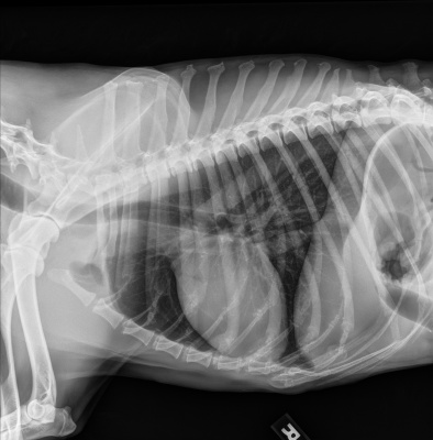
Charlie is a 10-year-old neutered male Wirehaired Pointer who presented to the IVS emergency clinic on July 26th for chronic weight loss over the past few months and anorexia. Over the past week, Charlie’s owner reported that he had been vomiting at night and again at the dog park a few days earlier. When Charlie’s owner contacted his regular veterinarian, they suggested dehydration and recommended increasing water intake. However, whenever Charlie drank water, he vomited it back up. Concerned, his owner brought him first to Urgent Vet, where abdominal radiographs revealed a possible gastrointestinal foreign body. Charlie was then referred to IVS for further treatment and care.
Physical Exam
On presentation, Charlie had a severely low body condition score (2/9). Abdominal palpation revealed marked pain and a palpable foreign object in the mid-ventral abdomen. His mucous membranes were pale pink and tacky, and periodontal disease was present. All other aspects of his physical exam were within normal limits.
Diagnostics
At Urgent Vet, abdominal radiographs showed a small intestinal obstruction caused by two mineralized foreign objects. At IVS, thoracic radiographs were taken to rule out metastatic neoplasia. IDEXX’s consult report indicated no intrathoracic evidence of metastatic disease.
A chemistry panel and complete blood count (CBC) were also performed. Results showed leukocytosis with severe neutrophilia, monocytosis, and eosinophilia, most consistent with inflammation and a possible stress leukogram. Mild hypocalcemia, hypochloremia, and hypoamylasemia were also noted, while all other values were within normal limits.
Diagnosis
Dr. Blake diagnosed Charlie with a gastrointestinal obstruction due to a foreign body versus neoplasia.
Treatment
The recommended treatment was an exploratory laparotomy. Prior to surgery, Charlie was hospitalized for fluid therapy, intravenous antibiotics (cefazolin), and pain management (methadone). Dr. Blake discussed potential surgical complications with the owner, who consented to proceed.
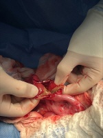
Charlie was started on a fentanyl CRI and pre-medicated with dexmedetomidine. He was induced with propofol and maintained on isoflurane anesthesia. During surgical preparation, his respiratory effort decreased. A ventral midline incision was made from sternum to pelvis. On entry, a section of jejunum containing the foreign body was exteriorized. Proximal to the obstruction, the intestines were adhered to the rectus abdominis. The affected intestinal segment was friable, ulcerated, and produced serosanguinous material on palpation. Several small adhesions were observed proximally, while the remainder of the gastrointestinal tract, liver, kidneys, and spleen appeared normal.
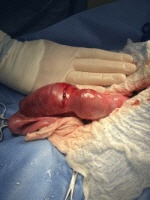
An enterotomy was performed, and hard pieces of hair and grass-covered plastic were removed. However, due to pressure necrosis and early perforation, the affected jejunal segment required resection and anastomosis. Charlie’s owner was contacted for consent, and Dr. Blake proceeded.
During surgery, Charlie’s pulses decreased and his respiratory effort became depressed. His gums appeared pale and tacky. Atipamezole was administered to reverse the dexmedetomidine. A 200 mL bolus of fluids was given, and fentanyl and isoflurane were discontinued, after which his vitals stabilized.
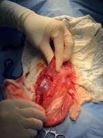
Doyen clamps were placed, the diseased jejunum was excised, and the disparity in lumen size was corrected by angling the transection. The jejunum was reconnected with simple interrupted sutures using absorbable material, beginning at the mesenteric and antimesenteric borders. A leak test was performed and was negative. The abdomen was lavaged with 2 L of warm saline before closure.
Post-operatively, Charlie was painful and required a fentanyl bolus, followed by a fentanyl CRI. Ketamine injections were available for additional pain control. Cefazolin continued every six hours, with pantoprazole and maropitant (Cerenia) added for acid reduction and anti-nausea effects. The following morning, antibiotics were switched to Unasyn.
At 9 a.m., Charlie showed no interest in food. He remained painful and vocalized throughout the day, requiring additional fentanyl and ketamine. By noon, he began eating, allowing transition from injectable to oral medications. He was discharged that evening on amoxicillin-clavulanate, maropitant, gabapentin, capromorelin (Entyce), omeprazole, and trazodone.
Gastrointestinal Obstructions
Gastrointestinal obstructions occur when indigestible or overly large material is ingested and cannot pass through the stomach or small intestine. Common canine culprits include toys, clothing, corn cobs, bones, and rocks, though other objects may also cause obstructions. In some cases, the foreign material causes intestinal perforation, leading to leakage of intestinal contents into the abdominal cavity.
Clinical signs often include vomiting, abdominal pain, diarrhea, dehydration, lethargy, and anorexia. If untreated, these patients can develop severe dehydration or sepsis, which may be life-threatening.
Treatment typically requires surgery, although endoscopy may be an option if the object remains in the stomach. Surgical approach depends on obstruction location: gastrotomy for the stomach or enterotomy for the small intestine. When intestinal segments are severely damaged, resection and anastomosis is required. Supportive therapy includes IV fluids, anti-nausea medications, multimodal pain relief, and perioperative antibiotics.
In resection and anastomosis, a healthy margin cranial and caudal to the diseased segment is identified. Mesenteric vessels supplying the affected segment are ligated, and the diseased intestine is removed. If lumen sizes differ, angled incisions are made to facilitate a better match before suturing. In Charlie’s case, the smaller lumen was cut at a 45° angle to match the markedly dilated proximal segment.
Patient Update
Charlie returned to IVS on August 7th for staple removal. His incision had healed, and the staples were removed successfully. He was discharged with the hope that he avoids foreign material ingestion in the future.
Sources
- Cohn LA, Côté E, eds. Côte's Clinical Veterinary Advisor: Dogs and Cats. 4th ed. Elsevier Mosby; 2020.
- Cornell University College of Veterinary Medicine. Gastrointestinal Foreign Body Obstruction in Dogs. Riney Canine Health Center. Published 2024. Accessed August 12th, 2025. https://www.vet.cornell.edu/departments-centers-and-institutes/riney-canine-health-center/canine-health-information/gastrointestinal-foreign-body-obstruction-dogs
- Ford RB, Mazzaferro EM, eds. Kirk & Bistner’s Handbook of Veterinary Procedures and Emergency Treatment. 9th ed. Elsevier Saunders; 2012.
- Mullen KM, Regier PJ, Ellison GW, Londoño L. The Pathophysiology of Small Intestinal Foreign Body Obstruction and Intraoperative Assessment of Tissue Viability in Dogs: A Review. Top Companion Anim Med. 2020;40:100438. doi:10.1016/j.tcam.2020.100438