
Gastrointestinal Foreign Body
Written by Torrin Kileen • 2025 Scholar
History
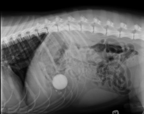
Ellie is a one-year-old, female spayed, Golden Retriever presenting for an endoscopic gastrointestinal foreign body removal. Ellie was transferred to Iowa Veterinary Specialty from Waukee-Clive rDVM after owners noticed lethargy and diarrhea for one day. The rDVM provided radiographs, bloodwork, and administered Cerenia, an antiemetic, to manage nausea. There appeared to be a rock present in the stomach on the abdominal radiographic images.
Physical Exam Findings & Diagnostics
Upon physical exam, Ellie had mild hyperthermia at 102.9 F and a slightly muddy mucous membrane. Her abdomen was tense on palpation indicating that she was likely uncomfortable. She was graded a 1/4 on the pain scale. Ellie had an otherwise unremarkable physical exam. Radiographs and bloodwork were forwarded from the rDVM, limiting the need for further diagnostics. It was recommended to repeat radiographs prior to the endoscopy to ensure the rock had not traveled further into the gastrointestinal tract where the foreign material could not be removed with an endoscope.
Diagnosis
Ellie was diagnosed with a gastrointestinal foreign body, suspected to be a rock on radiographs and confirmed/resolved with an endoscope performed by Dr. Bartlett.
Gastrointestinal Foreign Body Symptoms & Diagnostics
A gastrointestinal (GI) foreign body occurs when an animal ingests an object that is not digestible. Symptoms of a GI foreign body can vary depending on the object, where the object is within the GI tract, as well as when the animal ingests the object. Typical signs are vomiting, anorexia, abdominal pain, dehydration, diarrhea, or accumulation of abdominal fluid. Some objects such as fishhooks and sticks can cause perforations or holes in the GI tract, becoming life-threatening quickly, while others such as raw bread dough or rocks can cause obstructions that block food and water from passing.
Typically, for an animal presenting these symptoms, a veterinarian would recommend a complete blood count, serum chemistry, and abdominal radiographs. If these tests do not reveal an explanation for the symptoms, the radiographs may be repeated with barium contrast, or an ultrasound can provide additional information.
Treatment
There are a variety of treatment options based on the circumstances of the case. In Ellie’s case, radiographs were repeated at Iowa Veterinary Specialty prior to the endoscopy to ensure the rock remained in the stomach. As the rock had not yet passed into the intestines and was minimal risk for damaging tissue if removed endoscopically, that was the treatment plan. Ellie was placed under general anesthesia and an endoscope was used to visualize the stomach and proximal descending duodenum. The rock was found in the stomach, grasped with a wire basket attachment, and removed via the oral cavity.
More conservatively, gastrointestinal foreign bodies can be managed with supportive care, including fluids and monitoring for the object to pass on its own if it is small and safe enough. According to the National Library of Medicine, 80-90% of GI foreign bodies resolve without medical intervention. Inducing vomiting may be a viable treatment method if the material can safely pass through the esophagus. Sharp or caustic materials may cause more damage if removed with this method. If no conservative methods effectively remove the foreign body, it needs to be addressed surgically. This may be a gastrotomy or enterotomy (opening of the stomach or intestines) or a complete removal of damaged intestinal tissue, shortening the bowel.
While prognosis for this this condition is generally good, complications of treatments can occur. These include ileus (slowing or stopping of the intestinal tract), narrowing of the intestines, leaking into the abdomen, or short bowel syndrome. Short Bowel syndrome refers to when too much of the small intestine was damaged by the foreign body and needs to be removed during surgery. This condition typically occurs in cases exceeding 75% of the bowel removed and prevents proper absorption of nutrients.
Resources
- Gastrointestinal foreign bodies. American College of Veterinary Surgeons. (2025, June 4). https://www.acvs.org/small-animal/gastrointestinal-foreign-bodies/
- Jaan A, Mulita F. Gastrointestinal Foreign Body. [Updated 2023 Aug 28]. In: StatPearls [Internet]. Treasure Island (FL): StatPearls Publishing; 2025 Jan-. Available from: https://www.ncbi.nlm.nih.gov/books/NBK562203/
- Radiology. Surgical FAQ’s. (n.d.). https://www.senecafallsvet.com/radiology.pml