
Caudoventral Coxofemoral Luxation with a Closed Reduction
Written by Erica Kelly, First Year Veterinary Student • 2021 Scholar
Signalment and Presentation
Kevin, a 16-year-old neutered male domestic shorthair feline presented to IVS due to acute onset of left hind limb lameness.
History
Kevin’s owner noticed this afternoon that he was non-weight bearing on his hind left limb. His owner did not witness any traumatic events that may have caused an injury, however Kevin frequently jumps from windowsill to windowsill. Per Kevin’s owner, about 10 years ago he broke his hind left limb, but that has not caused any issues for Kevin since it first healed. No other concerns were mentioned by his owner.
Physical Exam
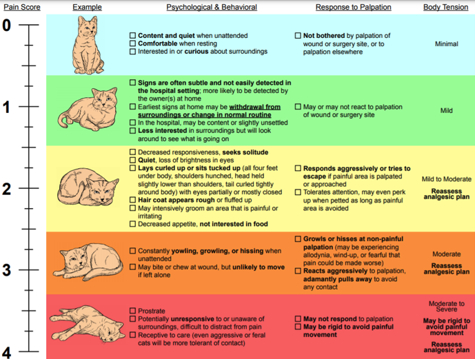
Upon physical exam, Kevin was BAR and neurologically appropriate. It was noted that he has iris degeneration in both eyes and severe periodontal disease. Kevin’s orthopedic exam revealed that he was non-weight bearing lame on his hind left limb, which he held abducted, and that he was painful on palpation of the femur/hip. It was determined that he had a pain score of 3 out of 4. There were no other significant exam findings at this time.
Colorado State University provides pain scoring scales to help tailor pain relief for each animal.
Diagnostics
Out of concern for an orthopedic injury, it was recommended that 3 view orthogonal orthopedic radiographs of the hips and stifles should be taken. Kevin’s owner consented, and he was given a pain control injection of hydromorphone prior to radiographs.
Radiographic Findings
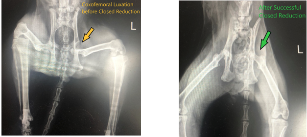
The left acetabulum is empty and the left femoral head is luxated ventrally and medially.
Diagnosis
Based on the radiographic findings, it was concluded that Kevin had a left femoral head luxation. The head of Kevin’s femur was displaced caudoventral to his pelvis. Coxofemoral luxations are one of the most common luxations in the dog and cat, with the majority of them being luxated craniodorsally – the opposite of Kevin’s luxation.
Treatment
There are four main treatment options for coxofemoral luxations including a closed reduction, open reduction, femoral head osteotomy (FHO), and hip replacement. Since Kevin’s trauma happened recently, he was a good candidate for a closed reduction followed by strict rest and pain medications. Closed reductions are most successful when attempted within the first 24-48 hours after the injury occurred and have a success rate of 50%.
In order to perform the closed reduction, Kevin would need to be placed under general anesthesia. Prior to this anesthetic event, we checked a blood chemistry to ensure he was a good candidate for anesthesia, and there were no concerning abnormalities noted.
Kevin was placed under general anesthesia and after two attempts, his coxofemoral luxation was successfully reduced via a closed reduction. Radiographs were taken to confirm that the reduction was successful. Kevin was sent home with buprenorphine – a pain medication, robenacoxib (Onsior) – an anti-inflammatory, and directions to endure strict rest until his recheck appointment in seven days.
Had Kevin’s closed reduction not been successful, an open reduction, FHO, or hip replacement would have been discussed. An open reduction is a surgical procedure in which the hip is surgically replaced and the supporting structures are restored. In most cases implants are placed to support the joint while it heals. This procedure has a very high success rate. In some patients a reduction is not possible due to poor hip conformation. In this case, a femoral head osteotomy (FHO), or hip replacement, would be pursued. FHOs are advised in patients that are less than 50 pounds, highly active dogs, and cats. A femoral head osteotomy involves producing a false joint by removing the femoral head and neck. In a false joint there is no bone to bone contact, but simply a joint capsule connecting two bones. Following this procedure, physical therapy is advised in order to maximize function of the joint. Performing an FHO eliminates the chance of reluxation.
References & Citations
- Hip Luxation, American College of Veterinary Surgeons https://www.acvs.org/small-animal/hip-luxation
- Coxofemoral Luxation: Tips for Closed Reduction, University of Illinois College of Veterinary Medicine https://vetmed.illinois.edu/coxofemoral-luxation-tips-for-closed-reductions/
- CSU Animal Pain Scales https://vetmedbiosci.colostate.edu/vth/services/anesthesia/animal-pain-scales/
- Coxofemoral Luxations in Cats. F. J. Pérez-Aparicio, Journal of Small Animal Practice, 1993, 34; 345-349 https://onlinelibrary.wiley.com/doi/epdf/10.1111/j.1748-5827.1993.tb02708.x
- Hip Dislocation in Dogs and Cats. Brooks, Wendy. 2005 https://veterinarypartner.vin.com/default.aspx?pid=19239&catId=102899&id=4952410