
Hemorrhagic Splenic Mass
Written by Nicole Clemen, 2nd year veterinary student • 2018 Scholar
History
Bodhi is a 10 year old intact male chocolate Labrador Retriever. He presented to IVS with lethargy, an inability to walk and labored breathing after an oncology appointment the day prior. He was weak and had to be carried into the clinic. Bodhi was transferred from oncology to emergency for appropriate treatment. The day prior during his oncology visit an ultrasound was performed and fine-needle aspirates were taken from his spleen and submitted for testing.
- Mucous membranes were tacky and pale pink
- Mandibular lymph nodes were enlarged
- Tachycardia (increased heart rate)
- Tachypnea (increased breathing rate)
- Positive fluid wave of abdomen (abdomen appears to have abnormal amounts of fluid within it, which is observable with a “wave” occurring with palpation and tapping of the sides of the abdomen)
- Depressed mood and difficulty standing
Once Bodhi was admitted, he had chest radiographs taken, an abbreviated abdominal ultrasound scan performed with fluid pulled from the abdomen, and blood work done which consisted of a complete blood count (CBC), chemistry, PT and PTT (clotting times), and he was hooked up to an ECG to monitor heart activity.
- Radiographs: the chest radiographs showed sternal lymphadenopathy (enlarged sternal lymph node), splenic neoplasia (abnormal mass attached to the spleen) and peritoneal effusion (presence of excess fluid in the abdomen)
- Ultrasound: ultrasound confirmed presence of small amounts of free fluid in the abdominal cavity, of which a sample was taken and a PCV obtained. The PCV was 40%.
- PCV, or packed cell volume, is a test that indicates the percentage of the blood that is red blood cells. This can indicate if an animal is anemic if the sample is obtained from the peripheral circulation, or if there is active bleeding into a body cavity, such as in this case. If the PCV of free fluid is high we know there is active bleeding.
- Blood work: Bodhi’s CBC and chemistry panel came back with elevated ALP, ALT, and white blood cell count. His peripheral PCV was 36%, which is mildly low, and his PT and PTT also came back normal.
- ALP (alkaline phosphatase) and ALT (alanine aminotransferase) are liver enzymes that can indicate liver disease and inflammation, but can sometimes also be elevated due to stress.
- The white blood cell count (WBC) is a reflection of the number of cells that generate immune responses in the body. An elevated WBC can indicate possible infection.
- PT (prothrombin time) and PTT (partial thromboplastin time) are tests that determine the ability of blood to clot by recording the length of time it takes to clot effectively and can detect the presence of a clotting disorder.
- ECG: Bodhi was having runs of VPCs, or ventricular premature contractions, meaning that is heart was contracting in an abnormal rhythm
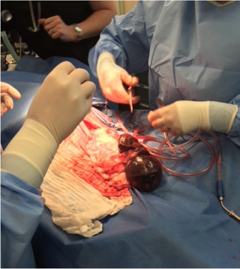
Abdominal bleeding from splenic mass(es), which were later discovered to be myelolipomas. An abdominal exploratory was performed resulting in a splenectomy (removal of the spleen). There were numerous nodules seen throughout the spleen on both the head and tail with one large, hemorrhaging lesion present on the head. Splenic hyperplasia (a mass present on the spleen due to increases in cell number) is not uncommon in dogs and can be harmless, however it is important to discern the cause of the changes as they could be due to neoplastic, or cancerous, changes. The spleen was submitted for testing through Iowa State University Pathology Department to determine the cellular basis of the mass and nodules on the spleen. The pathology report found mature adipocytes (fat cells) and hematopoietic tissue (tissue that produces red blood cells) together; this is collectively called a myelolipoma. Myelolipomas are typically benign. Evidence of hemangiosarcoma was not found. Hemangiosarcoma is a malignant cancer that frequently originates in the spleen in dogs and ought to be considered when a dog presents with masses and hemorrhaging of the spleen.
Due to the urgency of the situation, treatment was performed soon after admittance to the hospital. The owners opted to do an abdominal exploratory to discover the cause and stop the hemorrhage within the abdomen.
Treatment
Since Bodhi’s blood work came back without any significant and concerning news, surgery was begun as soon as he was ready in order to stop any continued bleeding. During surgery, a blood transfusion was also administered to compensate for blood loss that occurred prior to and during surgery. Prior to and after his surgery he was given a guarded prognosis. Bodhi was continuing to have VPC’s after surgery and was slow to recover. Twelve hours after surgery, his blood work showed a PCV of 29%, which is considered anemic, and he was also thrombocytopenic meaning his platelets were low at 0.3 per high powered field. Platelets are cell fragments that aid in clotting of blood, so low levels can indicate continued bleeding or that his body is not adequately recovering from surgery. He was hospitalized after surgery and placed on a HLK CRI, Cerenia, and IV fluids in addition to his ECG monitoring.
- A HLK CRI stands for hydromorphone, lidocaine and ketamine constant rate infusion. This essentially means that Bodhi has a constant infusion of hydromorphone, lidocaine and ketamine to sedate him and make him comfortable. Hydromorphone is a sedative and an analgesic. Lidocaine is effective at preventing irregular heart contractions, or arrhythmias, and also serves as an analgesic. Lastly, ketamine has analgesic effects as well. A CRI ensures that the optimal, desired amounts of these drugs are being delivered to tissues constantly and consistently.
- Cerenia is an anti-emetic drug, meaning that it reduces nausea and vomiting.
- IV fluids consist of isotonic solution of electrolytes that aids in replenishing body fluids lost from surgery or dehydration.
About twenty-four hours post-surgery, Bodhi was still having runs of VPC’s however they had decreased in frequency and are now considered sub-clinical, or unlikely to cause any negative side effects. He resumed eating and was stable. He was released from the hospital about a day and a half post-surgery. He was sent home on oral medications consisting of galliprant, codeine, and amoxi/clav:
- Galliprant is an NSAID that aids in controlling pain and inflammation.
- Codeine is also an oral analgesic to relieve moderate pain.
- Amoxi/clav stands for a combination of amoxicillin and clavulanate potassium which are both antibiotics that are used to treat urinary tract, skin and soft tissue infections.
Bodhi Today
Since his pathology report came back with good news that the masses on the spleen were most likely benign his treatment from here on out is much less involved, however rechecking of his platelets was advised in three to four days to see if they will return to normal levels. Normal platelet levels for dogs range from 8- 15 platelets per high powered field.
Since being released from the hospital, Bodhi has returned for a platelet rechecks twice in which his platelets went from 1.6 to 5, to 18 per high powered field, which is greatly improved. His incision was healing and he was doing well at home.
References & Citations
- Felizzola, C. (2016). Evaluation of Splenic Masses in Dogs: A Retrospective Study 2014/2015. Retrieved August 10, 2018 from https://www.vin.com/members/cms/project/defaultadv1.aspx?id=8311429&pid=20247&
- Plumb, D. C. (2005). Plumb's Veterinary Drug Handbook (8th ed.). Wiley Blackwell.
- Tilley, LP, Smith, FWK. Blackwell’s Five-Minute Veterinary Consult: Canine and Feline (6th Ed). Ames, Iowa: Wiley Blackwell.