Male Poodle - Vomiting, Anorexia, Increased Urination, and Increased Drinking
Written by Kayleen Hammer • 2021 Scholar
History
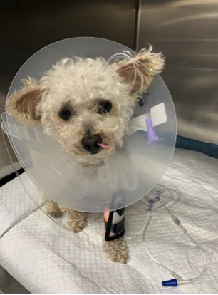
Rylee is a 7-year-old neutered male toy poodle that presented to Iowa Veterinary Specialties (IVS) for continued vomiting, anorexia, increased urination (polyuria), and increased drinking (polydipsia). Rylee went to the lake 6 days ago (July 4) with his owners and has been vomiting bile since then. Rylee was taken to his regular DVM 4 days ago (July 6) and prescribed Cerenia (an antiemetic) and Orbax (an antibiotic). However, Rylee continued to vomit while on Cerenia, and presented to the IVS emergency department on July 10.
Physical Exam-abnormal findings
- Tacky mucous membranes
- Normal mucous membranes are pink and moist. Mucous membranes are an indication of perfusion and hydration. Tackiness can indicate dehydration.
- Distended, tense abdomen with tympany.
- Tympany is a hollow percussion sound that can indicate an area is distended/filled with gas.
Differentials
Ileus
Ileus is the temporary cessation of intestinal muscle contractions that prevents food, fluids, GI secretions, and gas from flowing through the digestive tract. Ileus is not caused by a physical obstruction. Some causes of ileus are electrolyte imbalances, gastroenteritis due to infection, and drug effects.
Gastric foreign body/Pyloric Outflow Obstruction
Foreign bodies can also prevent ingesta from moving through the GI tract. If left untreated, this causes intractable vomiting that can lead to serious electrolyte imbalances, malnutrition, and aspiration pneumonia. The pylorus is a narrowing in the stomach orad to the duodenum that is a common site for obstruction to occur.
Diagnostics and Results
CBC (complete blood count): A CBC is done to evaluate the number and morphology of red blood cells, white blood cells, and platelets. In this case, we were particularly interested in the WBC to see if an infection/inflammatory response was occurring in Rylee.
Rylee’s CBC results:
- Leukocytosis (18490) Normal= 5050-16760
- Monocytosis (1664) Normal= 160-1120
- Neutrophilia (14792) Normal=2950-11640
Chemistry Panel:
A chemistry panel provides information on an animal’s overall health and organ function.
Rylee’s Chem panel results:
- Elevated BUN
- Elevated Globulin
These elevations are consistent with dehydration.
Abdominal Radiographs
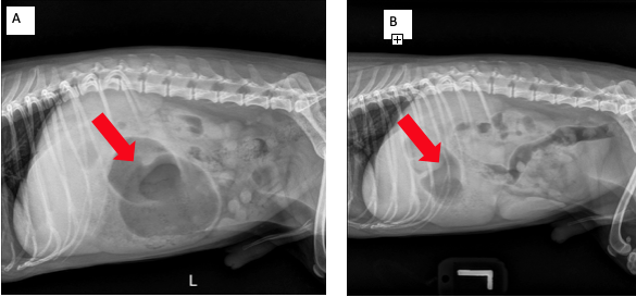
- This is one of the first radiographs taken shortly after Rylee’s arrival at IVS. The radiologist reported that Rylee’s stomach was moderately dilated, containing gas and fluid/soft tissue opacity. Gas is seen in the pylorus.
- Radiographs were taken later in the day to see if the obstruction was still present. Some foreign bodies pass on their own, and do not need surgery. However, in Rylee’s case, the stomach dilation and gas/soft tissue/fluid opacity remained after 12 hours of fasting. This indicates that Rylee needed surgery in order for his ingesta to properly flow through his gastrointestinal tract.

Diagnosis
Dr. Waller, a board-certified radiologist that reads all of the X-rays at IVS, diagnosed Rylee with
- Pyloric outflow obstruction, inflammatory gastritis
- Gastric foreign body: rounded, soft tissue gastric opacity
Treatment

July 10th
- Morning: After arriving at IVS, Rylee was given IV fluids, Cerenia IV, and Ondansetron IV (anti-nausea). A nasogastric tube was placed under sedation with butorphanol and propofol. Radiographs were taken (see A above). Aspiration of fluid was attempted to help determine if an obstruction was likely, no fluid was able to be suctioned. Rylee was NPO, meaning she couldn’t eat/drink by mouth.
- 1 pm: Rylee’s nasogastric tube was suctioned again, but no fluid was aspirated. At this point a foreign body was a strong concern.
- 8pm: Rylee remained NPO and repeat radiographs were taken (see B above). A foreign body was confirmed via radiographs.
July 11th
- 2 am: Rylee was premedicated, intubated, prepped and taken into surgery to remove his gastric foreign body. Dr. Blake made a 6cm ventral incision and evaluated the rest of the gastrointestinal tract. The small intestine and colon were normal. The stomach was distended and another incision was made between the greater and lesser curvatures of the stomach. Multiple chunks of potato were removed. The incision was closed and the stomach and abdomen were flushed with sterile saline. The body wall and subcutaneous layer were closed with separate simple continuous suture patterns. Staples were used to close the skin. Surgery took about 1 hour.
Outcome
Rylee recovered from surgery well and with no complications. Rylee was given fentanyl CRI for pain management and unasyn. Unasyn is a broad-spectrum antibiotic used to prevent bacterial infection. Rylee remained on fluid therapy, cerenia, and ondansetron. Rylee stayed an additional night until she was eating and drinking again. Today, Rylee is doing well and will hopefully stay away from potatoes!
References & Citations
- https://vcahospitals.com/know-your-pet/gastritis-in-dogs
- https://www.merckvetmanual.com/digestive-system/diseases-of-the-stomach-and-intestines-in-small-animals/gastrointestinal-obstruction-in-small-animals#:~:text=Pyloric%20stenosis%20can%20cause%20gastric,obstruction%20and%20are%20self%2Dlimiting.
- https://wagwalking.com/condition/pyloric-obstruction-stenosis


